Diatoms
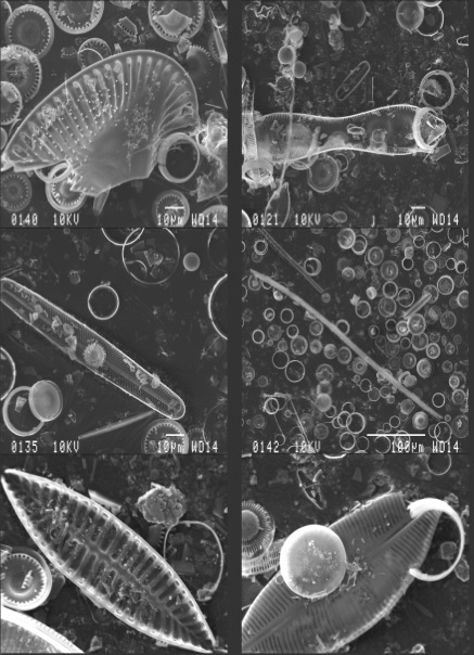
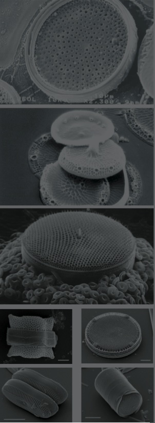
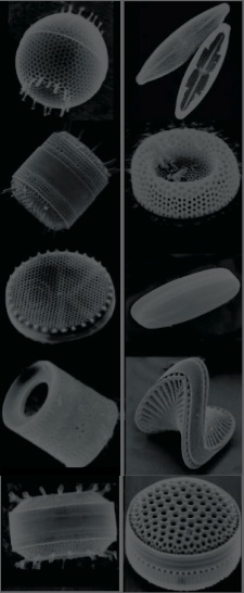
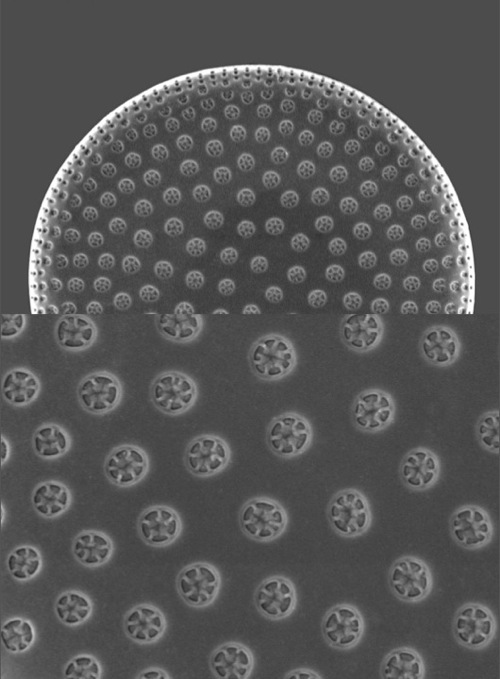
Diatoms are a major group of eukaryotic algae, and are one of the most common types of phytoplankton. Most diatoms are unicellular, although some form chains or simple colonies. A characteristic feature of diatom cells is that they are encased within a unique cell wall made of silica called a frustule. These frustules show a wide diversity in form, some quite beautiful and ornate, but usually consist of two asymmetrical sides with a split between them, hence the group name.
There are more than 200 genera of living diatoms, and it is estimated that there are approximately 100,000 extant species. Diatoms are a widespread group and can be found in the oceans, in freshwater, in soils and on damp surfaces. Most live pelagically in open water, although some live as surface films at the water-sediment interface (benthic), or even under damp atmospheric conditions. Although usually microscopic, some species of diatoms can reach up to 2 millimetres in length.
Most diatom species are non-motile but some are capable of an oozing motion. As their relatively dense cell walls cause them to readily sink, planktonic forms in open water usually rely on turbulent mixing of the upper layers by the wind to keep them suspended in sunlit surface waters. Some species actively regulate their buoyancy with intracellular lipids to counter sinking.
Diatoms cells are contained within a unique silicate (silicic acid) cell wall comprised of two separate valves (or shells). The biogenic silica that the cell wall is composed of is synthesised intracellularly by the polymerisation of silicic acid monomers. This material is then extruded to the cell exterior and added to the wall. Diatom cell walls are also called frustules or tests, and their two valves typically overlap one other like the two halves of a petri dish. In most species, when a diatom divides to produce two daughter cells, each cell keeps one of the two valves and grows a smaller valve within it. As a result, after each division cycle the average size of diatom cells in the population gets smaller. Once such cells reach a certain minimum size, rather than simply divide vegetatively, they reverse this decline by forming an auxospore. This expands in size to give rise to a much larger cell, which then returns to size-diminishing divisions. Auxospore production is almost always linked to meiosis and sexual reproduction. (source: Wikipedia)

Diatoms first appeared 200,000 million years ago, but flourished in Mesozoic times (ten million years ago) where they lived in such enormous numbers they left vast deposits of their shells up to three thousand feet thick. We call this diatomaceous earth and use it for filters, insulation, and as an abrasive.
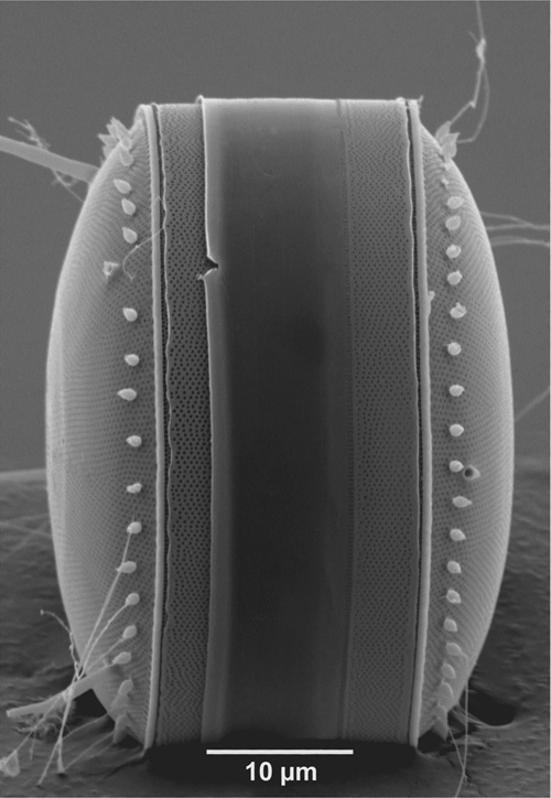
The glassy shell of a diatom comes in two parts, one fitting over the other exactly like a container with a lid.
The upper and lower halves are sculpted with exceedingly fine lines that actually are rows of ultra-microscopic pores through which the enclosed protoplasm remains in touch with the exterior environment.
Diatoms have been well studied both in their natural habitat and in cultures by biologists and there is therefore a wealth of knowledge on their biology and ecology. The protoplast of diatoms consists of a cytoplasmic layer that lines the interior of the frustule and surrounds a large central vacuole, within the cytoplasmic layer there is a diploid nucleus and two to several pigment-bearing plastids (the site of photosyntheseis). The diatom frustule is often likened to a pill-box or agar dish with an epitheca (larger upper valve), and a hypotheca (smaller lower valve). The vertical lip or rim of the epitheca is called the epicingulum, and the epicingulum fits over (slightly overlaps) the hypocingulum of the hypotheca. The epicingulum and hypocingulum with one or several connective bands make up the girdle.
(source: microscopy-uk.org.uk)